

B Cells
B lymphocytes (B cells) are an integral part of the humoral immune response due to their ability to produce antibodies against foreign antigens. B cells originate from hematopoietic stem cells (HSCs) in the bone marrow, which is seeded during embryonic development by HSCs from the fetal liver. The initial stages of B cell development are antigen-independent and involve the generation of several intermediary precursor cells that arise from B lymphocyte progenitor cells including Pre-pro-B cells, Pro-B cells, and Pre-B cells, which develop into immature B cells. During these stages of development, B cells undergo immunoglobulin gene rearrangement resulting in the expression of a mature B cell receptor (BCR) that is capable of binding to antigen. This is followed by a selection process that involves BCR editing or clonal deletion, designed to eliminate self-reactive immature B cells. The majority of immature B cells that survive this selection process leave the bone marrow and migrate to the spleen where they differentiate into transitional immature B cells that then become immunocompetent naïve mature B cells. Most naïve B cells develop into follicular B cells, while a small population becomes marginal zone B cells (frequently called IgM memory B cells in human). Following antigen-dependent activation, follicular B cells participate in germinal center reactions where they differentiate into memory B cells or long-lived, antibody-secreting plasma cells. While these developmental stages are similar for human B cells and mouse conventional B2 cells, a second mouse cell lineage (B1 cells) has also been described. B1 cells are abundant in the peritoneal cavities and can be further subdivided into B1a and B1b subsets. These along with other B cell precursor cells and functionally distinct subtypes including marginal zone B cells, follicular B cells, memory B cells, plasma cells, and regulatory B cells can be distinguished from each other based on the expression of specific cell surface and intracellular markers. R&D Systems and Novus Biologicals together offer the widest selection of fluorochrome-conjugated antibodies for detecting human and mouse B cell subsets and further characterizing these cells.

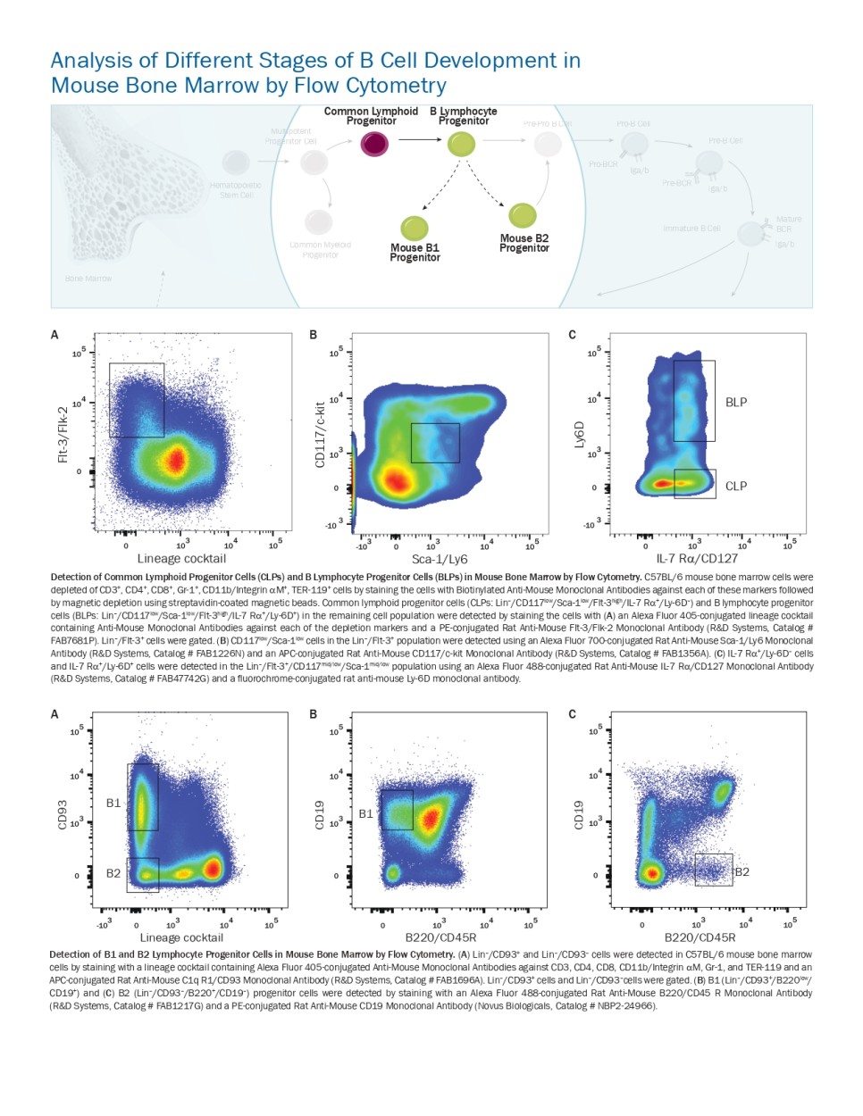
Analysis of Different Stages of B Cell Development in Mouse Bone Marrow by Flow Cytometry
Detection of Common Lymphoid Progenitor Cells (CLPs) and B Lymphocyte Progenitor Cells (BLPs) in Mouse Bone Marrow by Flow Cytometry. C57BL/6 mouse bone marrow cells were depleted of CD3+, CD4+, CD8+, Gr-1+, CD11b/Integrin aM+, TER-119+ cells by staining the cells with Biotinylated Anti-Mouse Monoclonal Antibodies against each of these markers followed
by magnetic depletion using streptavidin-coated magnetic beads. Common lymphoid progenitor cells (CLPs: Lin–/CD117low/Sca-1low/Flt-3high/IL-7 Ra+/Ly-6D–) and B lymphocyte progenitor
cells (BLPs: Lin–/CD117low/Sca-1low/Flt-3high/IL-7 Ra+/Ly-6D+) in the remaining cell population were detected by staining the cells with (A) an Alexa Fluor 405-conjugated lineage cocktail
containing Anti-Mouse Monoclonal Antibodies against each of the depletion markers and a PE-conjugated Rat Anti-Mouse Flt-3/Flk-2 Monoclonal Antibody (R&D Systems, Catalog # FAB7681P). Lin–/Flt-3+ cells were gated. (B) CD117low/Sca-1low cells in the Lin–/Flt-3+ population were detected using an Alexa Fluor 700-conjugated Rat Anti-Mouse Sca-1/Ly6 Monoclonal
Antibody (R&D Systems, Catalog # FAB1226N) and an APC-conjugated Rat Anti-Mouse CD117/c-kit Monoclonal Antibody (R&D Systems, Catalog # FAB1356A). (C) IL-7 Ra+/Ly-6D– cells
and IL-7 Ra+/Ly-6D+ cells were detected in the Lin–/Flt-3+/CD117mid/low/Sca-1mid/low population using an Alexa Fluor 488-conjugated Rat Anti-Mouse IL-7 Ra/CD127 Monoclonal Antibody
(R&D Systems, Catalog # FAB47742G) and a fl uorochrome-conjugated rat anti-mouse Ly-6D monoclonal antibody.
Detection of B1 and B2 Lymphocyte Progenitor Cells in Mouse Bone Marrow by Flow Cytometry. (A) Lin–/CD93+ and Lin–/CD93– cells were detected in C57BL/6 mouse bone marrow
cells by staining with a lineage cocktail containing Alexa Fluor 405-conjugated Anti-Mouse Monoclonal Antibodies against CD3, CD4, CD8, CD11b/Integrin aM, Gr-1, and TER-119 and an APC-conjugated Rat Anti-Mouse C1q R1/CD93 Monoclonal Antibody (R&D Systems, Catalog # FAB1696A). Lin–/CD93+ cells and Lin–/CD93–cells were gated. (B) B1 (Lin–/CD93+/B220low/
CD19+) and (C) B2 (Lin–/CD93–/B220+/CD19–) progenitor cells were detected by staining with an Alexa Fluor 488-conjugated Rat Anti-Mouse B220/CD45 R Monoclonal Antibody
(R&D Systems, Catalog # FAB1217G) and a PE-conjugated Rat Anti-Mouse CD19 Monoclonal Antibody (Novus Biologicals, Catalog # NBP2-24966).

Analysis of Different Stages of B Cell Development in Mouse Spleen by Flow Cytometry
Detection of Transitional 1 (T1) and Transitional 2 (T2) B Cells in Mouse Splenocytes. (A) C57BL/6 mouse splenocytes were stained with a PE-conjugated Rat Anti-Mouse CD19 Monoclonal Antibody (Novus Biologicals, Catalog # NBP2-24966) and CD19+ cells were gated. (B) B220+/CD43– cells in the CD19+ cell population were detected by staining with an Alexa
Fluor 750-conjugated Rat Anti-Mouse B220/CD45 R Monoclonal Antibody (R&D Systems, Catalog # FAB1217S) and an Alexa Fluor 488-conjugated Rat Anti-Mouse CD43 Monoclonal Antibody (Novus Biologicals, Catalog # NBP1-43413AF488). (C) Transitional 1 (T1) B cells (CD19+/B220+/CD43-/IgM+/IgDlow) and transitional 2 (T2) B cells (CD19+/B220+/CD43-/IgM+/
IgD+) were detected in the CD19+/B220+/CD43- population by staining with an Alexa Fluor 647-conjugated rat anti-mouse IgD monoclonal antibody and a PE-Cy7-conjugated Rat Anti-
Mouse IgM Monoclonal Antibody (Novus Biologicals, Catalog # NBP1-42940).
Detection of Transitional B1a and B1b Cells in Mouse Splenocytes. (A) C57BL/6 mouse splenocytes were stained with an Alexa Fluor 488-conjugated Rat Anti-Mouse CD43 Monoclonal Antibody (Novus Biologicals, Catalog # NBP1-43413AF488) and an Alexa Fluor 594-conjugated Rat Anti-Mouse CD23/Fce RII Monoclonal Antibody (R&D Systems, Catalog # FAB6900T). CD43+/CD23– cells were gated. CD1dmid cells in the CD43+/CD23– population were detected by staining with an Alexa Fluor 700-conjugated Rat Anti-Mouse CD1d Monoclonal Antibody
(Novus Biologicals, Catalog # NBP1-43461AF700; Data not shown). (B) Transitional B1a cells (CD5+/CD19high/CD1dmid/CD23–/CD43+) and transitional B1b cells (CD5–/CD19high/CD1dmid/
CD23–/CD43+) were detected in the CD1dmid/CD23-/CD43+ population by staining with a PE-conjugated Rat Anti-Mouse CD19 Monoclonal Antibody (Novus Biologicals, Catalog # NBP2-
24966) and an APC-conjugated Rat Anti-Mouse CD5 Monoclonal Antibody (R&D Systems, Catalog # FAB115A).

Detection of Marginal Zone and Follicular B-2 Cells in Mouse Splenocytes. (A) C57BL/6 mouse splenocytes were stained with a PE-conjugated Rat Anti-Mouse CD19 Monoclonal Antibody (Novus Biologicals, Catalog # NBP2-24966) and an Alexa Fluor 488-conjugated Rat Anti-Mouse CD43 Monoclonal Antibody (Novus Biologicals, Catalog # NBP1-43413AF488). CD19+/ CD43– cells were gated. (B) Follicular B-2 cells (CD19mid/CD1dmid/CD23+/CD21low/CD43–) and marginal zone B-2 cells (CD19mid/CD1dhigh/CD23–/CD21high/CD43–) were detected in the
CD19+/CD43– population by staining with a fl uorochrome-conjugated anti-mouse CD21 monoclonal antibody and an Alexa Fluor 700-conjugated Rat Anti-Mouse CD1d Monoclonal
Antibody (Novus Biologicals, Catalog # NBP1-43461AF700). CD1dmid/CD21low and CD1dhigh/CD21high cells were gated. (C) Follicular B-2 cells (CD1dmid/CD21low) and marginal zone B-2 cells
(CD1dhigh/CD21high) were stained for CD21 and CD23 using a fl uorochrome-conjugated anti-mouse CD21 monoclonal antibody and an Alexa Fluor 594-conjugated Rat Anti-Mouse CD23/
Fce RII Monoclonal Antibody (R&D Systems, Catalog # FAB6900T).
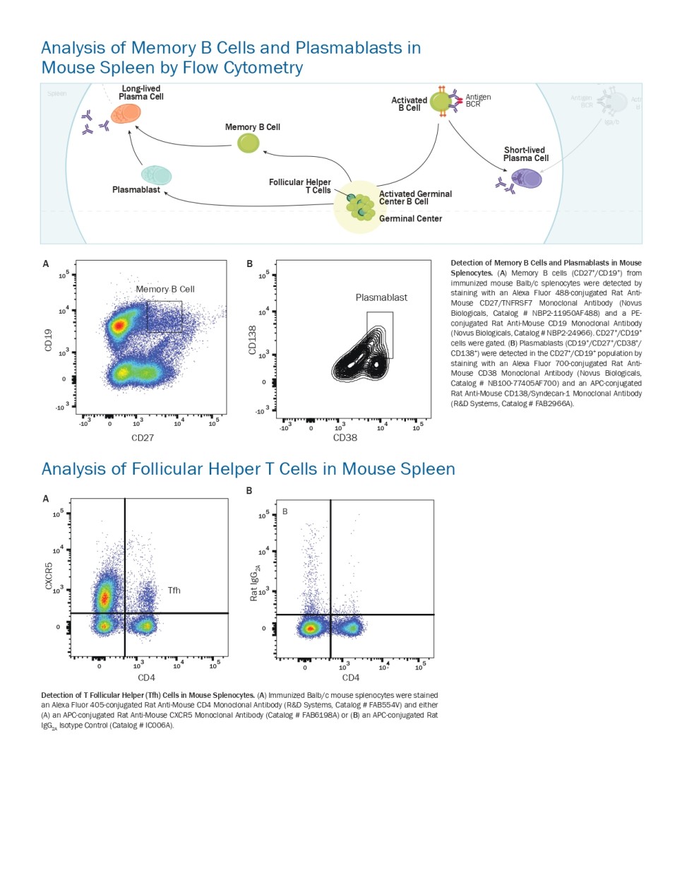
Analysis of Memory B Cells and Plasmablasts in Mouse Spleen by Flow Cytometry
Detection of Memory B Cells and Plasmablasts in Mouse Splenocytes. (A) Memory B cells (CD27+/CD19+) from
immunized mouse Balb/c splenocytes were detected by staining with an Alexa Fluor 488-conjugated Rat Anti- Mouse CD27/TNFRSF7 Monoclonal Antibody (Novus Biologicals, Catalog # NBP2-11950AF488) and a PE- conjugated Rat Anti-Mouse CD19 Monoclonal Antibody (Novus Biologicals, Catalog # NBP2-24966). CD27+/CD19+
cells were gated. (B) Plasmablasts (CD19+/CD27+/CD38+/
CD138+) were detected in the CD27+/CD19+ population by
staining with an Alexa Fluor 700-conjugated Rat Anti- Mouse CD38 Monoclonal Antibody (Novus Biologicals, Catalog # NB100-77405AF700) and an APC-conjugated Rat Anti-Mouse CD138/Syndecan-1 Monoclonal Antibody (R&D Systems, Catalog # FAB2966A).
Analysis of Follicular Helper T Cells in Mouse Spleen
Detection of T Follicular Helper (Tfh) Cells in Mouse Splenocytes. (A) Immunized Balb/c mouse splenocytes were stained an Alexa Fluor 405-conjugated Rat Anti-Mouse CD4 Monoclonal Antibody (R&D Systems, Catalog # FAB554V) and either (A) an APC-conjugated Rat Anti-Mouse CXCR5 Monoclonal Antibody (Catalog # FAB6198A) or (B) an APC-conjugated Rat IgG2A Isotype Control (Catalog # IC006A).

Analysis of Human Plasma Cells and Memory B Cells by Flow Cytometry
Detection of Plasma Cells and Memory B Cells in Human Peripheral Blood Mononuclear Cells by Flow Cytometry. Plasma cells (CD3–/CD19low/CD20–/low/CD38high/BLIMP1+) in human peripheral blood mononuclear cells were detected by staining
with (A) an Alexa Fluor 405-conjugated Mouse Anti-Human CD3e Monoclonal Antibody (R&D Systems, Catalog # FAB100V) and an Alexa Fluor 594-conjugated Mouse Anti-Human CD19 Monoclonal Antibody (R&D Systems, Catalog # FAB4867T). CD3–/CD19+ cells were gated. (B) Expression of CD20/MS4A1 and CD38 on cells in the CD3–/CD19+ gate was determined
by staining with an APC-conjugated Mouse Anti-Human CD20/MS4A1 Monoclonal Antibody (R&D Systems, Catalog # FAB4225A) and a PerCP-conjugated Mouse Anti-Human CD38 Monoclonal Antibody (R&D Systems, Catalog # FAB2404C). CD20–/low/CD38+ cells were gated. (C) Expression of BLIMP1 in the CD20–/low/CD38+ cell population was determined by
staining with a PerCP-conjugated Mouse Anti-Human CD38 Monoclonal Antibody (R&D Systems, Catalog # FAB2404C) and a PE-conjugated Mouse Anti-Human BLIMP1/PRDM1 Monoclonal Antibody (R&D Systems, Catalog # IC36081P). (D) Memory B cells were also detected in the starting population of human peripheral blood mononuclear cells by staining with a Fluorescein-conjugated Mouse Anti-Human CD27/TNFRSF7 Monoclonal Antibody (R&D Systems, Catalog # FAB382F) and an Alexa Fluor 594-conjugated Mouse Anti-Human CD19 Monoclonal Antibody (R&D Systems, Catalog # FAB4867T)
Detection of Pax5 Expression in Human B Cells by Flow Cytometry. Human peripheral blood mononuclear cells were surface stained using an APC-conjugated Mouse Anti- Human CD19 Monoclonal Antibody (R&D Systems, Catalog # FAB4867A). Cells were then fi xed and permeabilized and stained intracellularly using a Rabbit Anti-Human Pax5 Monoclonal Antibody (R&D Systems, coming soon) followed by a PE-conjugated Goat Anti-Rabbit Antibody (R&D Systems, Catalog # F0110).


Additional Products for B Cell Research
In addition to the large selection of fluorochrome-conjugated antibodies that are offered by R&D Systems and Novus Biologicals, R&D Systems offers several kits to simplify your B cell research including B Cell Isolation and Expansion Kits, and recombinant proteins for B cell culture and differentiation. We also offer a wide range of assays designed to provide insight into the B cell-mediated immune response such as B Cell ELISpot Development Modules, Luminex® Immunoglobulin Isotyping Assays, and ELISA Kits to quantify B cell-secreted molecules.
Luminex® Mouse Immunoglobulin Isotyping Assay
This bead-based assay is designed to simultaneously quantify multiple mouse immunoglobulin isotype concentrations in cell culture supernatants, serum, or EDTA plasma. The assay is designed for use with the Luminex MAGPIX®,
Luminex 100™, Luminex 200™, or Bio-Rad® Bio-Plex® dual laser, flow-based sorting and detection analyzers.

B Cell Expansion Kit R&D Systems® CellXVivo™ Human B Cell Expansion Kit contains all of the reagents necessary
to expand a population of 107 human B cells 3–5-fold.

'RD_Brochure' 카테고리의 다른 글
| 3-D Matrices & Assay Kits (0) | 2021.06.14 |
|---|---|
| DuoSet® ELISA Development Systems (0) | 2021.06.14 |
| Products for Wnt Research (0) | 2021.06.14 |
| Myeloid-derived Suppressor Cells (0) | 2021.06.14 |
| Epithelial to Mesenchymal Transition (0) | 2021.06.14 |


